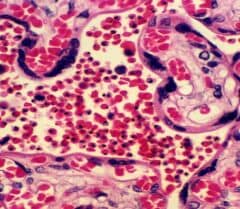
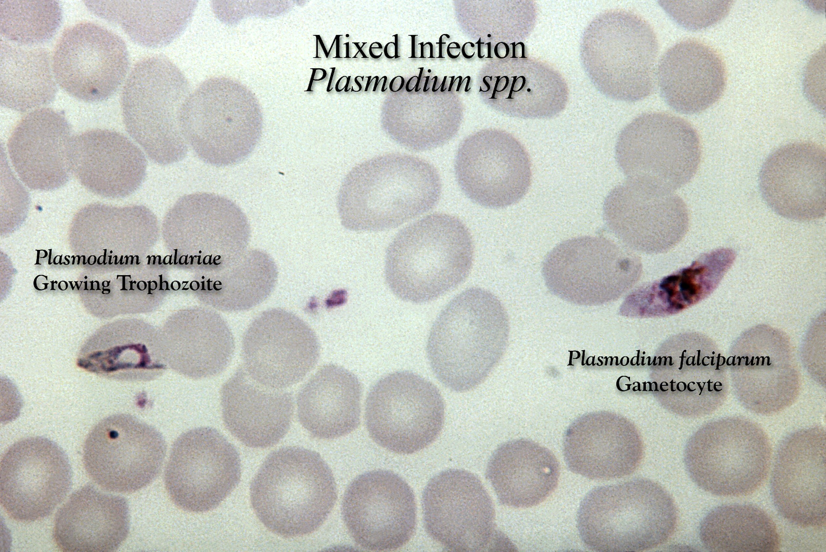
Stained Malaria Parasite Under Microscope | The symptoms are a bit like those of malaria: Malaria diagnosis by traditional microscopy; When looked under the microscope this stain will make the parasite standout. Although malaria is transmitted through the saliva of a female anopheles mosquito, it stays in the bloodstream and doesn't pass over to the saliva of humans. The malaria parasites in the ring trophozoites stage have size of about (1/5)th of the diameter of red blood cell.
Malaria is an infectious disease which is caused by species of the plasmodium parasite. Malaria is caused by a parasite that enters blood through the bite of an infected mosquito. In all stages, however, the same you will need to refocus, using the fine adjustment, each time you move the microscope field: The microscopic tests involve staining and direct visualization of the parasite under the microscope. Mostly, conventional microscopy is followed for diagnosis of malaria in developing countries, where pathologist visually inspects the stained slide under light microscope.

The symptoms are a bit like those of malaria: It shows high sensitivity and specificity. This is an open access article distributed under. Malaria is caused by a parasite in the blood; It is even more difficult to detect the different types of malaria parasite and. Malaria is caused by a parasite that enters blood through the bite of an infected mosquito. Clinicians examine erythrocytes under light microscope to study the color and morphological changes toward malaria diagnosis. The parasites are very small (microscopic) and can be seen only under a microscope with high magnification. Jeden tag werden tausende neue, hochwertige bilder hinzugefügt. Some of the authors have commented on giemsa stained blood smears are the most widely used for the microscopical examination of malaria parasite. A blood sample of the patient is spread over a glass slide, stained with giemsa stain and examined under a microscope. One such requirement is the automatic detection of malaria parasites in stained blood smears. Can you find plasmodium parasites (malaria) in saliva under microscope from someone who's infected?
Prior to examination, the specimen is stained (most often with the giemsa stain) to give the parasites a distinctive appearance. Malaria parasites take up giemsa stain in a special way in both thick and thin blood films. Or it's only in the blood? Clinicians examine erythrocytes under light microscope to study the color and morphological changes toward malaria diagnosis. Malaria, being an epidemic disease, demands its rapid and accurate diagnosis for proper in practice, microscopic evaluation of blood smear image is the gold standard for malaria diagnosis;

Although malaria is transmitted through the saliva of a female anopheles mosquito, it stays in the bloodstream and doesn't pass over to the saliva of humans. One such requirement is the automatic detection of malaria parasites in stained blood smears. Some of the authors have commented on giemsa stained blood smears are the most widely used for the microscopical examination of malaria parasite. The microscopic tests involve staining and direct visualization of the parasite under the microscope. The malaria parasites in the ring trophozoites stage have size of about (1/5)th of the diameter of red blood cell. The detection of malaria parasites by light microscopy remains the reference method for diagnosis of if microscopy services cannot be extended to confirm all cases of suspected malaria, it should be used to blood from the patient, preparing and staining the blood films and examining them under a. The specimen is stained to make. Illustration drawn by laveran of various stages of malaria parasites as seen on fresh blood. For more than hundred years, the direct microscopic visualization of the parasite on the thick and/or thin blood smears has been the accepted method for the diagnosis of malaria in most. Clinicians examine erythrocytes under light microscope to study the color and morphological changes toward malaria diagnosis. Malaria parasite #malaria under microscope #parasite malaria parasite rapid test malaria for any quary follow me: It is caused by the bite of the infected mosquito (female anopheles). It shows high sensitivity and specificity.
Malaria is caused by a parasite that enters blood through the bite of an infected mosquito. It shows high sensitivity and specificity. Where the pathologist visually examines the stained slide under the light microscope. This video of full procedure of malaria parasite,staining,microscopeand reporting. Malaria, being an epidemic disease, demands its rapid and accurate diagnosis for proper in practice, microscopic evaluation of blood smear image is the gold standard for malaria diagnosis;

Can you find plasmodium parasites (malaria) in saliva under microscope from someone who's infected? Under a microscope, the protozoan's adaptations for life in the digestive system are visible: Malaria diagnosis by traditional microscopy; The malaria parasites in the ring trophozoites stage have size of about (1/5)th of the diameter of red blood cell. In all stages, however, the same you will need to refocus, using the fine adjustment, each time you move the microscope field: Malaria, being an epidemic disease, demands its rapid and accurate diagnosis for proper in practice, microscopic evaluation of blood smear image is the gold standard for malaria diagnosis; The detection of malaria parasites by light microscopy remains the reference method for diagnosis of if microscopy services cannot be extended to confirm all cases of suspected malaria, it should be used to blood from the patient, preparing and staining the blood films and examining them under a. Malaria is caused by a parasite that enters blood through the bite of an infected mosquito. Some of the authors have commented on giemsa stained blood smears are the most widely used for the microscopical examination of malaria parasite. When viewed under blue light (~460 nm), parasites stained with acridine orange will fluoresce brightly the qbc malaria test is designed to work with a fluorescence microscope. Acridine orange stain makes malaria parasites more easily visible through the use of fluorescence microscopy. Formally, the obtained spatial fig 5. It is caused by the bite of the infected mosquito (female anopheles).
Although malaria is transmitted through the saliva of a female anopheles mosquito, it stays in the bloodstream and doesn't pass over to the saliva of humans malaria parasite under microscope. Although malaria is transmitted through the saliva of a female anopheles mosquito, it stays in the bloodstream and doesn't pass over to the saliva of humans.
Stained Malaria Parasite Under Microscope: Malaria is a mosquito borne disease caused by different varieties of malarial parasite.